Eating Disorders in the Brain: What Neuroimaging Is Teaching Us
Eating disorders are often thought of in terms of discernible symptoms like eating behaviors and relationships to food and body. But beneath the more-visible symptoms lies a deeper, less obvious story playing out inside the brain. As neuroimaging techniques have advanced, researchers are uncovering real, measurable differences in brain structure, function, and connectivity in people who experience eating disorders. This article explores what neuroimaging is teaching us about eating disorders: how they may affect the brain, how brain biology may contribute to risk, what it means for treatment and recovery, and why this perspective matters.
If you or a loved one is navigating an eating disorder, you are not alone. Understanding the neurology of eating disorders can help with gaining insight and finding a path toward healing.
Eating Disorders and the Brain
An eating disorder is a complex mental health condition that affects how a person thinks about food, body image, and self-worth. Eating disorders can involve restricting food, binge eating, purging, compulsive exercise, or a combination of these behaviors. The most well-known types include anorexia nervosa, bulimia nervosa, and binge eating disorder, but there are also other forms, such as avoidant/restrictive food intake disorder (ARFID) and other specified feeding or eating disorders (OSFED). Regardless of type, eating disorders are serious illnesses that can affect anyone, regardless of age, gender, body size, body weight, or background. They are influenced by a mix of factors including genetics, brain biology, personality traits, trauma, cultural pressures, and life experiences. Over time, eating disorders can take a serious toll on both physical and emotional health, impacting everything from heart function to relationships. Yet, with early intervention, compassionate care, and comprehensive treatment, full recovery is possible.
There are many existing diagnostic tools and treatments for eating disorders. Increasingly, researchers are looking to neuroimaging to help understand why and how eating disorders develop, and how treatment options can be improved. Neuroimaging provides a window into the brain’s structure and function, asking questions such as:
- Are there measurable differences in the brains of people with eating disorders versus those without?
- Do those differences change with recovery or treatment?
- Can neurobiology help explain why some symptoms (such as restriction, bingeing, purging, distorted body image) persist or recur?
Neuroimaging can help move the conversation beyond known eating disorder behaviors and symptoms toward a fuller understanding of the brain, body and mind together.
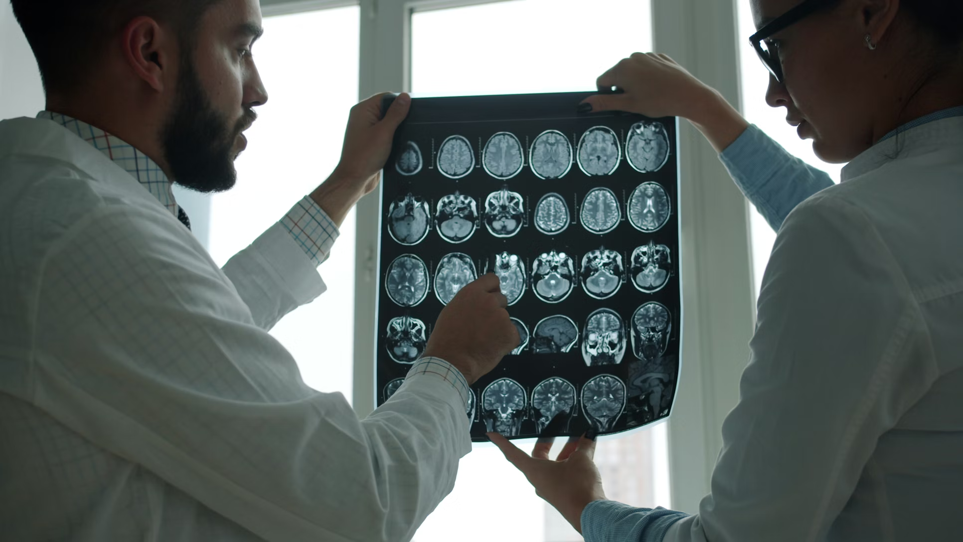
What Is Neuroimaging?
Before diving into findings, it helps to understand what neuroimaging is. Neuroimaging, a neuroscience technology, refers to a group of medical brain imaging techniques used to visualize and study the structure and function of the brain and central nervous system. It provides detailed insights into the brain’s anatomy, metabolism, and activity, aiding in the diagnosis and understanding of various neurological and psychiatric conditions. Some common types of neuroimaging include:
- Structural MRI (Magnetic Resonance Imaging): This technique captures the anatomy of the brain, visualizing how big different regions are, the thickness of cortex, and volume of sub-regions.
- Functional MRI (fMRI): fMRI measures brain activity by detecting changes in blood flow while the participant is either at rest or completing a task.
- Resting-state fMRI (rs-fMRI): This is a form of fMRI that looks at connectivity between brain networks when the person is not performing a specific task.
- Diffusion Tensor Imaging (DTI): DTI measures characteristics of the brain’s wiring and connectivity between regions.
- Positron Emission Tomography (PET) / SPECT: Though less common in recent eating disorder research, PET technology is used to examine metabolism and neurotransmitters.
These tools allow researchers to investigate three broad areas: brain structure, brain function, and brain connectivity. Each offers insights into how eating disorders might manifest at the neural-system level, which can help healthcare professionals better understand and treat eating disorders.
EDs and Brain Structure
Research into structural brain changes for those experiencing eating disorders has revealed a number of consistent patterns. It is important to emphasize that many of these findings are correlational, which means researchers cannot always say whether the brain structure causes the behavior or vice versa. Nonetheless, the findings are meaningful.
Cortical thickness, brain volume, and white-matter
The brain is a vulnerable organ. One study noted that food restriction, such as in the case of anorexia nervosa, as well as bingeing and purging behaviors are associated with lower regional brain volumes or thinner cortical thickness, though some changes may reverse with weight restoration. In other words, the brain may show structural shrinkage or thinning during periods of illness. This may reflect the biological stress of malnutrition and altered metabolism.
Sub-regions and brain circuits
Studies have found reductions in volume in regions important for body-image perception, interoception (feeling one’s internal bodily state), and cognitive control for those experiencing eating disorders. For instance, in a systematic review of bulimia nervosa and binge eating disorder, researchers concluded that there were reductions and increases across a range of areas, including brain regions important for self-regulation and visual/spatial processing. This could result in dysfunction in feedback between the brain and body.
Recovery and plasticity
Encouragingly, some neuroimaging studies have found that many structural changes appear to reverse with treatment and eating disorder recovery. This suggests the brain is plastic and responsive to treatment and that early intervention matters. The degree of recovery can depend on the duration and severity of the eating disorder, another reason the neurological perspective underscores urgency in treatment.
Brain Function and Eating Disorders
Moving from anatomy to activity brings us deeper into how the brain processes food, body image, reward, and social cues in the context of eating disorders.
Reward processing and food cues
One of the most studied areas of neuroimaging is the brain’s reward circuitry. In people with eating disorders, especially binge eating disorder and bulimia nervosa, studies show altered responses to food cues. For example, one study found that individuals who binge eat display lower dopamine release, changes in volume of brain regions related to impulse control, and altered frontostriatal connectivity, which controls reward processing. Simply put, the brain’s desire for food may be dysregulated for those experiencing eating disorders. This may help explain why binge eating can feel compulsive or escape-like rather than simply driven by hunger.
Interoception, body-image, & self-perception
Brain regions involved in sensing internal body states, self-referential thinking, and the neural network that prioritizes importance show altered activation in those who struggle with eating disorders. A recent study of body-image distortion found changes in the amygdala, insula, anterior cingulate cortex, and connectivity among parietal/temporal/occipital regions. This suggests that when someone with an eating disorder looks in the mirror, thinks about food, or senses fullness/hunger, there may be a different brain response compared to someone without an eating disorder.
Resting-state network connectivity
When participants simply rest in a scanner, studies show altered connectivity in key brain networks for those experiencing eating disorders. Those include:
- The default-mode network (DMN): associated with self-referential thinking, mind-wandering.
- The salience network (SN): detects and filters relevant stimuli.
- The central executive network (CEN): involved in planning, cognitive control.
For example, a review of fMRI-based eating disorder research found that in those with anorexia nervosa, there was reduced connectivity in areas linked to facial and social cognition, and increased connectivity in regions tied to aesthetic judgment and social anxiety. Participants with binge eating disorder showed diminished connectivity within the salience network, and increased connectivity in the default-mode network. These findings imply that even when resting, the brains of people with eating disorders may be wired differently, and potentially predisposed to certain cognitive or emotional states.
So what?
From these structural and functional findings, several key insights emerge, each with implications for how we think about prevention, treatment, and recovery.
Reducing stigma
The neuroimaging evidence reinforces what clinicians and loved ones know: eating disorders are not about willpower. The brain displays measurable changes in structure and function, whether those changes are caused by, contribute to, or result from the disorder. Recognizing the brain basis can help reduce stigma and encourage compassion. Knowing that there are real brain changes associated with eating disorders can shift the conversation away from judgement-based sentiments and toward a network of support and care.
Everybody’s different
It’s important to note that brain findings vary widely based on several factors: type of eating disorder, condition severity, stage of recovery, and individual factors. A universal neurological profile does not exist. Differences in brain function and structure will influence how eating disorder treatment impacts an individual and what recovery can look like. This underscores the importance of personalized treatment approaches.
Hope for recovery
One of the most hopeful findings in research around neuroimaging and eating disorders is that many structural brain alterations improve with recovery. With the help of a care team, interventions like psychiatry, therapy, and individualized dietary plans, recovery of both the body and brain is possible. That being said, brain changes are greater with more severe or prolonged conditions. The earlier someone gets help, the better chance to minimize the negative impact on the brain and maximize neurological recovery. Early intervention and sustained care can make a difference from the inside out.
Additionally, these findings encourage a system of holistic care in eating disorder recovery. Neurological changes don’t exist in isolation. They interact with psychological, social, and behavioral factors. A treatment plan that includes nutrition, therapy, body-image work, family support, and medical monitoring is essential. Working closely with an eating disorder care team can help you find your unique, holistic path toward recovery.

The Future of Neuroimaging and Recovery
As neuroimaging continues to advance, its greatest promise may lie not just in revealing what’s different in the brain, but in helping people reach more sustainable recovery. In the coming years, brain-scanning tools may help clinicians see how recovery takes root, not only in eating behaviors but also in the gradual rewiring of thought patterns, emotions, and self-perception. This knowledge could guide more holistic, person-centered care, bridging the biological, psychological, and social aspects of eating disorders.
Imagine being able to show someone that their brain is becoming stronger and healthier as they nourish themselves and reconnect with their body; that recovery is not only about the visible changes to one’s body and behaviors, but also about the brain itself learning balance, flexibility, and calm. Researchers are also exploring how neuroimaging might be used alongside mindfulness, somatic therapies, and other integrative approaches to help people tune in to their internal cues and rebuild trust in their bodies. The future isn’t about replacing therapy with technology, it’s about deepening our understanding of how body, brain, and mind work together. By bringing compassion and science into closer partnership, neuroimaging can help with finding a path toward recovery.
The neuroimaging revolution in eating-disorder research is not just academic. It has real implications for how we understand, communicate, treat, and recover from these serious conditions. The brain is central: it is where hunger meets emotion, body-image meets self-identity, and behavior meets biology. The story of eating disorders is not only about food, weight, or image, it is about how the body and brain work together.
For the National Alliance for Eating Disorders, our mission remains: to provide support, information, resources, and hope. Knowing what is happening in the brain adds a powerful dimension to that mission, reinforcing that eating disorders are treatable conditions where early help, comprehensive care, and supportive communities make a difference.
Seek Help
If you or a loved one is experiencing an eating disorder, you are not alone. Recovery is possible and help is available with the National Alliance for Eating Disorders.